In routine, non–fat-suppressed spin-echo MRI, CSF is usually dark on T1-weighted images and bright on T2-weighted images. Fat often appears bright on T1. These are useful clues, not a way to identify every series: FLAIR, fat suppression, gadolinium, and other sequence choices can change the appearance.
T1 vs T2 MRI: the practical comparison
T1- and T2-weighted images emphasize different tissue relaxation properties. The comparisons below describe typical, non–fat-suppressed spin-echo or fast spin-echo images—not every possible MR sequence.
| Feature | T1-weighted | T2-weighted |
|---|---|---|
| CSF / simple fluid | Usually dark (low signal) | Usually bright (high signal) |
| Fat | Commonly bright | Variable; often remains fairly bright without fat suppression |
| Edema / increased free water | Often relatively low signal | Often high signal; typically easier to appreciate on fluid-sensitive images |
| Typical use | Anatomy, tissue characterization, and pre-/post-contrast comparison | Fluid-sensitive assessment, including edema and many pathologic processes |
| Classic spin-echo timing | Short TR + short TE | Long TR + long TE |
Signal is relative, not a fixed color assigned to each tissue. A T2-weighted image is not automatically a fat-suppressed image.
Why T1 and T2 produce different contrast
T1 describes how longitudinal magnetization recovers after excitation. T2 describes how transverse magnetization decays as spins lose phase coherence through spin–spin interactions. Both processes happen in tissue after excitation; they are not separate scanner types.
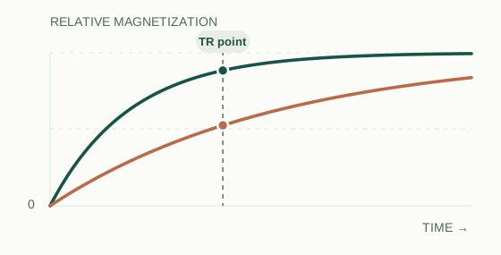
Read the marker: By the indicated TR, short-T1 tissue has recovered more. A short TR can emphasize this difference in conventional spin-echo imaging.
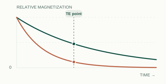
Read the marker: By the indicated TE, long-T2 tissue retains more transverse signal. A longer TE can emphasize this difference.
How TR and TE influence weighting
In a conventional spin-echo sequence, TR (repetition time) controls how much longitudinal magnetization has recovered before the next excitation. A shorter TR makes differences in T1 recovery more influential. TE (echo time) sets how long transverse magnetization has decayed by the time the echo is sampled. A longer TE makes differences in T2 decay more influential.
This is a framework for conventional spin echo, not a universal lookup table for every modern sequence. Fast spin echo, inversion recovery, and gradient-echo acquisitions can involve additional parameters, including echo trains, inversion time (TI), and flip angle. Do not assign a weighting from TR and TE alone when the sequence is unfamiliar.
How to recognize T1 and T2 on a brain MRI
Start with the series name and acquisition details. Then use anatomy as a cross-check—rather than guessing from the brightest structure.
- Check the CSF. In routine brain imaging, dark ventricular and sulcal CSF favors T1 weighting; bright CSF favors T2 weighting.
- Check fat and white matter. On a typical T1-weighted brain image, scalp fat tends to be bright and white matter is often brighter than gray matter. On conventional T2-weighted images, the white–gray matter contrast commonly reverses.
- Look for suppression or preparation. Dark CSF on T2-FLAIR does not make it a T1 image. A fat-suppressed T1 sequence will not show the usual bright fat.
- Confirm the series label. Check whether the acquisition is SE, FSE/TSE, GRE, FLAIR, STIR, or another sequence before interpreting its contrast.
Why an MRI protocol often includes both
T1-weighted imaging often provides clear anatomic detail and is central to evaluating signal changes after gadolinium administration. Enhancement is assessed by comparing appropriately matched pre- and post-contrast T1-weighted acquisitions; a single bright spot is not, by itself, proof of enhancement.
T2-weighted imaging is sensitive to changes in water content. It can make edema, fluid collections, and many lesions more conspicuous, but high T2 signal is not specific to any one diagnosis. A complete study uses several complementary sequences to answer a clinical question.
Four exceptions worth remembering
T2-FLAIR
T2-sensitive contrast with CSF deliberately suppressed. Ventricular CSF may look dark even when abnormal water-rich tissue remains bright.
Fat suppression
Fat can be dark on T1- or T2-weighted images when a suppression technique is applied. Always read the sequence label.
STIR
STIR applies an inversion pulse and chooses an inversion time (TI) to null signal from tissue with a particular T1—usually fat. It is not chemically selective like spectral fat suppression; tissues with similar T1 values may also be suppressed, including some enhancing tissues after contrast.
Gradient echo / T2*
Gradient-echo contrast depends on factors including flip angle, TR, TE and susceptibility-related T2* decay. Spin-echo TR/TE rules alone are not enough to identify or interpret every GRE image.
T1 and T2 MRI: frequently asked questions
Is fluid always white on T2 MRI?
No. Simple fluid is typically bright on routine T2-weighted spin-echo images, but FLAIR suppresses CSF, and other sequence choices can change fluid signal.
Is fat dark on T2-weighted MRI?
Not necessarily. Fat can remain relatively bright on many T2-weighted fast spin-echo images. It becomes dark when appropriate fat-suppression methods are used.
Is T1 or T2 better for finding a lesion?
Neither is universally better. T2-sensitive images can highlight water-related changes, while T1 and post-contrast T1 images add different information. The protocol depends on the clinical question.
Are T1 and T2 images the same as T1 and T2 maps?
No. Conventional weighted images mix several contrast influences; quantitative T1 or T2 mapping is a separate measurement approach.